

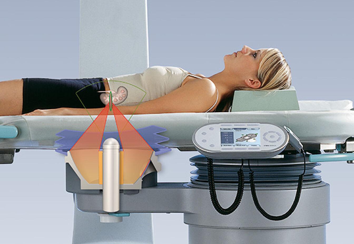
One of the area of advancement in ESWL is the refinement of imaging techniques. Real-time imaging is crucial during Extracorporeal Shock Wave Lithotripsy procedures to accurately locate the stone and ensure the shock waves are properly targeted. Traditionally, fluoroscopy, which uses X-rays, was used for stone visualization. However, the development of ultrasound and computed tomography (CT) scanning has provided superior imaging capabilities with reduced radiation exposure. Ultrasound allows for real-time monitoring of the stone position and movement during the procedure, while CT scanning provides detailed three-dimensional images for precise stone localization and targeting.
Additionally, the use of dual-energy CT scanning has further improved stone characterization. Dual-energy CT can differentiate between different types of stones based on their composition, such as calcium-based stones, uric acid stones, or cystine stones. This information helps urologists tailor the ESWL settings and treatment approach to optimize stone fragmentation and clearance.
Read More- https://cmiblogdailydose.blogspot.com/2023/06/extracorporeal-shock-wave-lithotripsy.html