

How does a Colposcope Work?
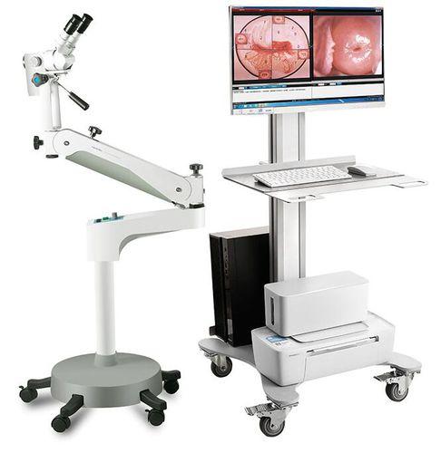
A colposcope consists of an illuminated magnifying lens system and filters that can display tissue surfaces in various wavelengths of light. The physician places a speculum into the vagina to gently hold the walls apart and provide access to the cervix. They then use the colposcope, which is mounted on an adjustable arm, to visually inspect the cervix. Filters allow visualization of vascular patterns and subtle tissue changes using techniques like acetic acid application and iodine staining. A camera attachment can record and document findings as well. Targeted biopsy forceps are used to collect samples from any suspicious lesions for histopathological examination.
Applications of Colposcopy in Early Cancer Detection
Colposcopy plays a vital role in cervical cancer screening programs worldwide. Abnormal Pap test results indicating possible precancerous lesions or malignancies prompt a Colposcope examination. This non-invasive procedure allows close-up inspection of the transformation zone, where most cervical cancers originate. Colposcopic findings help determine if further testing or treatment is needed, such as loop electrosurgical excision procedure (LEEP) or cone biopsy for high-grade lesions and early cancers. Accurate colposcopic assessment is key to ensuring prompt evaluation and management of abnormalities that could potentially progress to invasive cancer if left unaddressed.
Get More Insights on Colpscope