

Western Blotting Wonders: Expanding Frontiers in Research
Introduction to Western Blotting
Western obscure, also known as immunoblotting, is a laboratory technique used to detect specific proteins in a sample of tissue homogenate or extract. It uses gel electrophoresis to separate native or denatured proteins by the length of the polypeptide chains. After electrophoresis, the proteins are transferred to a membrane (usually nitrocellulose or PVDF) where they are probed with antibodies specific to the target protein. Enzyme-linked secondary antibodies are used to detect and visualize the bands of interest on the membrane.
Sample Preparation and Gel Electrophoresis
The first steps involve preparing a cellular or tissue extract containing the protein of interest. Cells or tissues are lysed to release intracellular contents, and debris is removed by centrifugation. The protein concentration of the cleared lysate is determined using a colorimetric or fluorometric assay. An aliquot of the sample containing equal amounts of total protein is then mixed with a denaturing protein loading buffer containing SDS and reagents to reduce disulfide bonds.
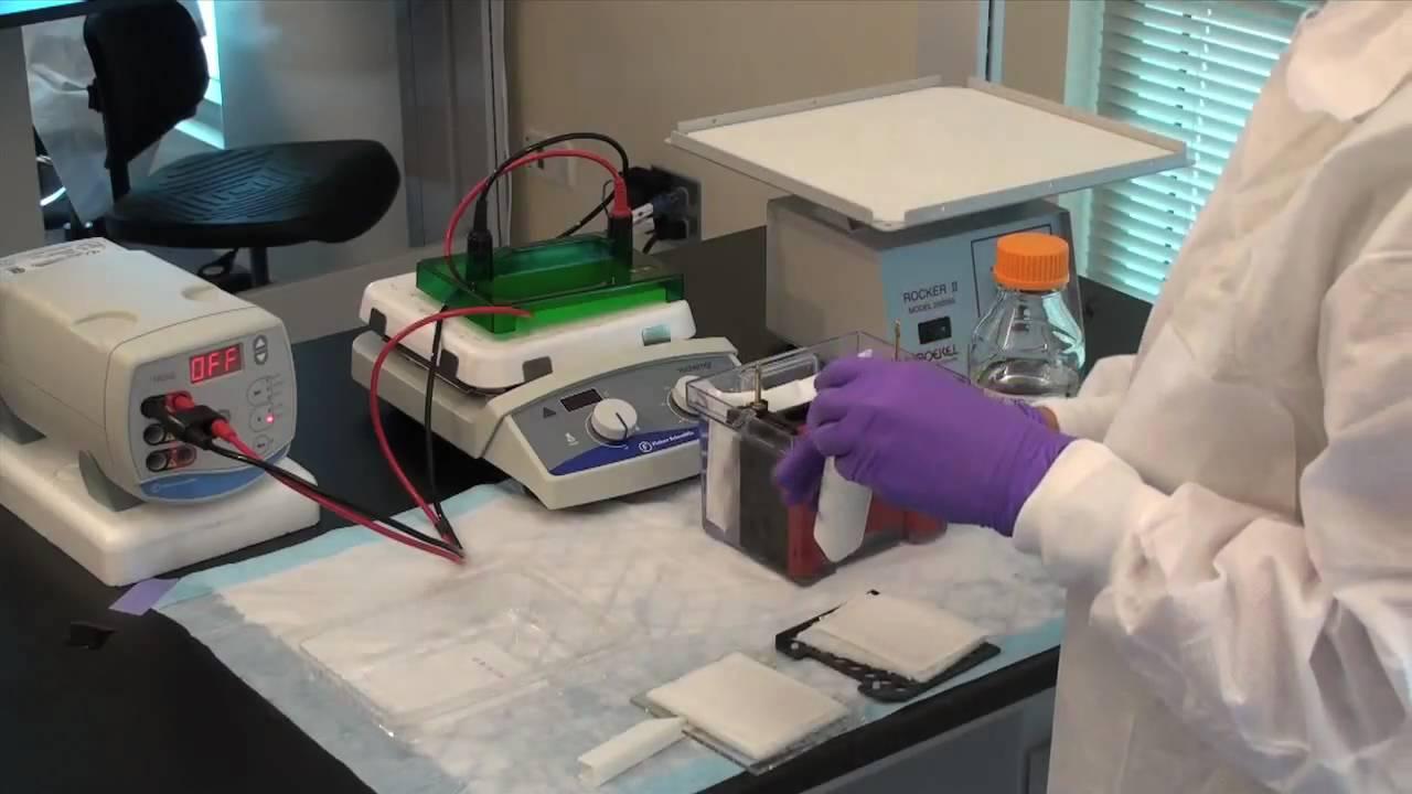
The prepared protein samples are loaded onto a polyacrylamide gel alongside known molecular weight protein standards. Gel electrophoresis is used to separate proteins based on their molecular weights as they migrate through the gel in response to an electric current. For Western obscure, proteins are resolved on SDS-PAGE gels containing SDS, which denatures the native structure and gives each protein a uniform negative charge proportional to its mass.
Transfer of Proteins to Membrane
Following electrophoresis, the separated proteins must be transferred from the gel onto a stable matrix for detection. Wet electrotransfer presses the proteins from the gel onto a nitrocellulose or PVDF membrane in a process called blotting. The membrane acts as a solid support for further processing and detection steps. Efficient transfer is confirmed by Ponceau S staining of the membrane to visualize total protein patterns. The remaining sites on the membrane are then blocked to prevent non-specific antibody binding.
Primary and Secondary Antibody Incubation
The membrane is incubated with a primary antibody specifically targeted against the protein of interest. This primary antibody binds directly to any transferred protein on the membrane that matches its target epitope. Unbound primary antibody is washed away before applying a species-specific secondary antibody linked to an enzyme like horseradish peroxidase. The secondary antibody binds to the primary antibody adhered to the protein band to form an antibody-antigen-antibody "sandwich."
Substrate Incubation and Detection
After further washing, the membrane is incubated with a luminescent substrate optimized for the enzyme used. The enzymatic reaction produces light in proportion to the amount of bound secondary antibody and target protein present in each band. Chemiluminescent, fluorescent or chromogenic substrates are commonly used depending on the desired detection system. The target protein band is visualized as a discrete band on X-ray film or CCD camera for analysis.
Applications and Considerations for Western Blotting
Western obscure has become an invaluable technique in molecular and cell biology due to its high specificity and sensitivity. It can detect protein targets from picogram to nanogram levels depending on antibody choice. Common applications include analyzing protein expression levels, post-translational modifications, protein-protein interactions, antibody validation, and signaling pathway activity.
However, there are also limitations to consider with Western obscure. Protein abundance does not always correlate with activity level. Quantitative measurements require densitometric analysis and normalization to loading controls. Antibody specificity needs to be carefully validated. Molecular weight markers are an approximation, and protein migration can vary with experimental conditions. Proper experimental design and controls are crucial for obtaining reproducible and biologically meaningful results from Western blots.
Optimization of Western obscure Protocol
Several parameters may be adjusted to improve Western blot results depending on the proteins and antibodies used:
- Gel concentrations, pore size and running conditions influence protein separation and transfer efficiency.
- Membrane material, transfer buffer composition, voltage and time impact successful protein transfer from gel to membrane.
- Blocking reagent, concentration, and duration prevent nonspecific antibody binding to the membrane.
- Primary and secondary antibody concentrations, incubation periods, and buffers are optimized for signal detection without background noise.
- Washing conditions between antibody bindings remove unbound antibodies for clear band visualization.
- Substrate choice depends on the detection system available, and incubation conditions influence sensitivity of detection.
With practice and troubleshooting, the Western blot protocol can be optimized to sensitively and reliably detect target proteins of interest. Careful attention to controls and standards facilitates quantification and validation of results.
In summary, Western obscure is a critical technique for protein detection that combines the resolution of gel electrophoresis with specific antibody-based identification. Through transfer and probing protocols, it allows immunochemical identification and analysis of proteins isolated from complex biological systems. With proper optimization and execution, Western obscure remains an invaluable tool in molecular and cell biology research.