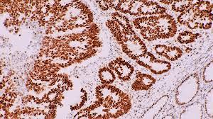
Immunohistochemistry (IHC) is a technique that is used to detect antigens (proteins) in cells of a tissue section by exploiting the principle of antibodies binding specifically to antigens in biological tissues. The detection of antigens relies on the use of specific antibodies that are usually linked to an enzyme or a fluorescent dye. The principal of IHC relies on the specificity of the interaction between antigens and their corresponding antibodies. By combining this specificity with a detection system, IHC allows visualizing specific proteins in tissues and cells.
Antibodies Used in Immunohistochemistry
IHC employs highly specific antibodies that are capable of recognizing antigens or proteins of interest within cells and tissues. Typically, monoclonal antibodies are used in IHC as they are generated against single epitopes, resulting in highly specific antibody binding. The antibodies used can be either primary or secondary. Primary antibodies are those that are specific for the targeted antigen. Secondary antibodies bind primary antibodies and are usually linked to an enzyme, fluorochrome or other molecules that provide the means for antigen detection. Commonly used secondary antibodies include anti-mouse or anti-rabbit IgG antibodies coupled to horseradish peroxidase (HRP), alkaline phosphatase or fluorescent dyes.
Antigen Retrieval in Immunohistochemistry
Antigen retrieval is a key pre-treatment step conducted before IHC staining to make antigens more accessible to the antibodies by reversing formaldehyde-induced cross-linking. Formaldehyde is commonly used to fix tissues during sample processing and this causes antigens to become masked by cross-linking to other proteins and components in tissues. Antigen retrieval treatments employ heatinduced epitope retrieval using various buffers, pH conditions and temperatures to break the protein cross-links and retrieve or uncover the antigen epitopes. This increases the number of antigenic sites available for antibody binding, thereby enhancing signal intensity and staining quality. Popular antigen retrieval methods involve microwave or pressure cooker heating of tissue sections immersed in buffer solutions like citrate or EDTA. The choice of pH conditions and buffer depends on the nature of antigen.
Detection Systems Used in Immunohistochemistry
Once primary antibodies have specifically bound to target antigens within the tissue sample, detection of antigen-antibody binding can be achieved using enzyme conjugated secondary antibodies along with a chromogenic substrate or a fluorophore conjugated secondary antibody.
- Enzyme-linked detection: Secondary antibodies conjugated to enzymes like HRP or alkaline phosphatase are used. When substrate for the enzyme is added, a color precipitate forms indicating sites of antigen-antibody binding. Common HRP substrates are diaminobenzidine (DAB) or aminoethyl carbazole (AEC) which yield brown or red precipitate respectively.
- Fluorescent detection: Secondary antibodies conjugated to fluorochromes like fluorescein isothiocyanate (FITC) or cyanine dyes are employed. When exposed to specific wavelengths of light, the labeled sites emit light of a characteristic color allowing visualization of antigen localization under a fluorescent microscope. Compared to enzyme labeling, fluorescence offers superior sensitivity and multiplexing capability.
Controls in Immunohistochemistry
Various control experiments are performed along with IHC staining to validate specificity and accuracy of the procedure. Negative controls involve omission of the primary antibody to establish that any staining observed is due to specific antibody-antigen interactions. Isotype controls employing primary antibodies of the same species and subclass but with irrelevant specificity assess non-specific secondary antibody staining. Positive controls use a tissue known to express the antigen of interest to ensure procedure is working satisfactorily. Serial section and blocking peptide controls check staining reproducibility. Proper controls determine the true immunoreactivity of a specimen and avoid false interpretation of staining results.
Applications of Immunohistochemistry
IHC has widespread applications in research, clinical diagnosis and forensics. It allows localization of antigens in both healthy and diseased tissues giving valuable insights into physiological and pathological processes. In research, IHC assists protein expression profiling, stem cell identification and developmental studies. Clinically, IHC helps diagnose infections, classify tumors, establish prognosis and predict therapy response through immunophenotyping. IHC also aids forensic identification of cells, tissues and biofluids by detecting antigens unique to tissue/cell types. Emerging applications involve automated quantitative IHC analysis and multiplexing techniques for co-detection of several antigens simultaneously in a sample. With continuous development of novel antibodies and detection methods, IHC plays a significant role as an indispensable technique in biomedical sciences.
Conclusion
In summary, this is a powerful technique that exploits antigen-antibody binding for visualizing specific proteins in tissues and cells. Combined with various detection systems, IHC provides exquisite localization of antigens at single cell level resolution. With appropriate antibody validation and control experimentation, IHC serves as an invaluable research and diagnostic tool across many areas of biomedical sciences. Advances in molecular techniques and automation of IHC are expected to drive its applications further in both research and clinical domains.
Priya Pandey is a dynamic and passionate editor with over three years of expertise in content editing and proofreading. Holding a bachelor's degree in biotechnology, Priya has a knack for making the content engaging. Her diverse portfolio includes editing documents across different industries, including food and beverages, information and technology, healthcare, chemical and materials, etc. Priya's meticulous attention to detail and commitment to excellence make her an invaluable asset in the world of content creation and refinement.
(LinkedIn- https://www.linkedin.com/in/priya-pandey-8417a8173/


