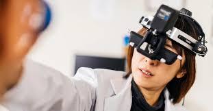
Ophthalmoscopes: A Valuable Medical Device for Examining the Eye

The ophthalmoscope was invented in 1851 by German physician Hermann von Helmholtz. Helmholtz designed the first functional one, which consisted of a light source and a set of adjustable lenses that could be held close to the eye. This allowed doctors to view the inside of the eye for the first time and examine structures like the retina. Early versions were challenging to use and lacked brightness, but Helmholtz's basic design proved the starting point for modern versions of the instrument.
Improvements continued throughout the latter half of the 19th century. Optometrist William Wilde and American surgeon George Tiffany both developed devices featuring improved lighting from kerosene lamps in the 1850s. In the 1890s, Joseph Leiter incorporated multiple reflectors driven by compressed air to provide brighter illumination. This electric device became a standard in eye exams.
Workings
All modern devices operate on similar principles. They focus a bright light into the pupil and provide magnification for the user to view inside structures. A basic one contains:
- A light source - Typically LED lights now replace older incandescent bulbs. Battery powered for portability.
- Lenses - Converging lenses project the light beam into the eye. Diopter scales allow adjustment for near- or far-sightedness.
- Mirror - A 45 degree mirror deflects the light beam into the eye at an oblique angle for a clear view behind the pupil.
- Magnification - Lenses in the viewing head can range from non-magnifying to 20x magnification for close examination.
To perform an exam, the lit end of the device is held close to the patient's eye while the physician looks through the viewing lens. Direct and indirect observation techniques allow inspecting the optic disc, vessels, macula and peripheral retina. Comparison to diagrams aids in detecting lesions or abnormalities.
Examination With an Ophthalmoscope
A standard eye exam with an device has several key steps:
1. Pupil Dilation - Drops may be applied to dilate the pupil for a clearer view of the retina. This allows more light into the back of the eye.
2. Disc Evaluation - The optic disc where retinal vessels join is examined. Changes in color, size or anatomy indicate issues like glaucoma.
3. Vessels - Retinal arteries and veins are inspected for narrowing, bulging, copper wiring, blood clots or aneurysms.
4. Macula - This central retina responsible for sharp, straight-ahead vision is checked for drusen, holes or other macular degeneration signs.
5. Peripheral Retina - The edges of the retina are viewed to detect detachments, tears or abnormalities. Diabetes can cause proliferation in these areas.
6. Pressure - An attached tonometer measures intraocular pressure, a key test for glaucoma screening.
Beyond these basic steps, doctors may use additional lenses, techniques or dyes during exams depending on a patient's history or symptoms discovered. A device remains essential for non-invasive evaluation of the inner eye.
Advances in Ophthalmoscope Technology
Significant improvements continue to be made to enhance these and their clinical usefulness. Some notable developments include:
- Indirect devices - A small mirror permits observation from the side rather than straight through for an upright retinal image.
- Wide-Angle or Panoramic Lenses - Provide a wider view up to 150-200 degrees rather than standard 30-45 degrees.
- Digital devices - Toric lenses capture retinal photos and video for comparison over time or remote consulting.
- Scanning Laser devices - Use multiple low-power lasers to obtain high-resolution digital images of the retina.
- Hybrid Contact Lens Models - Incorporate lenses directly into eyepieces for hands-free examination and imaging.
- Artificial Intelligence Integration - AI can compare images to databases and help detect common retinal diseases automatically.
As technology continues advancing eye care, devices remain a fundamental tool empowering physicians worldwide to screen for sight-threatening conditions and monitor patients' eye health. Their importance shows no sign of diminishing as medicine progresses.
So in summary, the device opened new possibilities for examining the inner eye upon its development in the 1800s. Constant refinements since have made it a mainstay diagnostic device, and emerging technologies promise to further strengthen its clinical utility long into the future. As a gateway to the retina, this invention holds great significance.
Priya Pandey is a dynamic and passionate editor with over three years of expertise in content editing and proofreading. Holding a bachelor's degree in biotechnology, Priya has a knack for making the content engaging. Her diverse portfolio includes editing documents across different industries, including food and beverages, information and technology, healthcare, chemical and materials, etc. Priya's meticulous attention to detail and commitment to excellence make her an invaluable asset in the world of content creation and refinement.
(LinkedIn- https://www.linkedin.com/in/priya-pandey-8417a8173/
- Art
- Causes
- Crafts
- Dance
- Drinks
- Film
- Fitness
- Food
- Games
- Gardening
- Health
- Home
- Literature
- Music
- Networking
- Other
- Party
- Religion
- Shopping
- Sports
- Theater
- Wellness
- IT, Cloud, Software and Technology


