


Multiple Organ System Tissue Microarrays for Histology Researchers
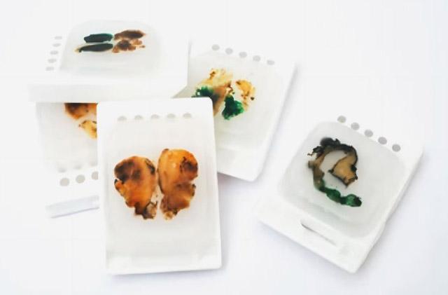
CD BioSciences, a US-based biotechnology company focusing on the development of imaging technologies, is proud to introduce its new Multiple Organ System Tissue Microarrays for histology researchers to study a wide range of biological processes and diseases across multiple organ systems. These microarrays provide a powerful tool for comprehensive and efficient high-throughput analysis of different organ systems.
Tissue microarray (TMA) helps to study a large number of tissue samples in a single section. In this technique, hundreds to thousands of tissue samples can be extracted from different paraffin blocks and precisely placed in a single recipient paraffin block. TMA helps to amplify the resource material and provides uniform staining conditions without affecting the original donor block. It is faster and less expensive than traditional staining methods and facilitates immunocytochemistry (IHC), fluorescence in situ hybridisation (FISH), in situ hybridization (ISH) or RNA analysis in a single section of a recipient block.
Multi-organ system tissue microarrays are slides containing tissue samples from several different organs in the body that allow researchers to simultaneously analyse protein expression or other molecular markers from different organ systems using immunohistochemistry or other staining techniques, providing a high-throughput method to study disease progression or tissue-specific differences in different organs on a single platform. By consolidating diverse biological information into a compact format, researchers can streamline their pathogenesis studies and accelerate the pace of biomarker discovery and drug development.
CD BioSciences now provides comprehensive Multiple Organ System Tissue Microarrays for histology researchers, e.g., histology technicians, histopathologists, and other research professionals, to facilitate the study of biological processes and diseases across multiple organ systems. These microarrays are meticulously constructed using advanced techniques to ensure optimal tissue preservation and efficiency and enable researchers to efficiently analyze and compare multiple tissues simultaneously, saving time and resources.
For example, the Digestive System Tumor Tissue Microarray (Catalog NO. MOCT060) comprises 140 cases, each containing 80 cores representing 6 distinct tumor types found in the digestive system. Ten cases were analyzed for each tumor type: esophagus squamous cell carcinoma, gastric adenocarcinoma, colon adenocarcinoma, rectum adenocarcinoma, hepatocellular carcinoma, and pancreas adenocarcinoma. These cases were compared to their respective normal adjacent tissues. Additionally, the microarray included normal tissues from the esophagus, stomach, colon, rectum, liver, and pancreas, with 2-5 cases analyzed for each type, totaling 7 cases.
The Multiple Organs Tumor and Normal Tissue Microarray (Catalog NO. MOCT078) contains 40 cases each of five common cancers: lung non-small cell carcinoma, prostate adenocarcinoma, breast invasive ductal carcinoma, colon adenocarcinoma, and pancreas duct adenocarcinoma. To provide a comparative baseline, the microarray also includes 10 samples of normal tissue for each of the lung, prostate, colon, and pancreas, along with 7 samples of normal breast tissue and 3 samples of adjacent normal breast tissue. Each case is represented by a single core. This microarray is applicable to IHC, ISH and other routine histology procedures.
CD BioSciences is committed to providing researchers with the highest quality tools to study complex biological processes. For more information about the Multiple Organ System Tissue Microarrays, please visit https://www.bioimagingtech.com/multiple-organ-system-tissue-microarrays.html.
About CD BioSciences
CD BioSciences is a biotechnology company committed to the development of imaging technology for many years. Its scientists can utilize high-content imaging, nanoparticle imaging, imaging flow cytometry, time-lapse imaging, and other techniques to image cell structure, cell migration, cell proliferation, pathogen infection mechanisms, and interactions between protein molecules.

 English
 Arabic
 Français
 Español
 Português
 Deutsch
 Türkçe
 Dutch
Dutch
 Russian
 Romaian